Skip to main content
List
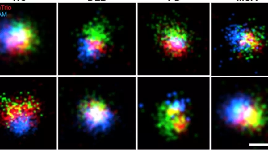
Kavli Oxford researchers reveal differences in alpha-synuclein associated with neurodegenerative diseases
20 October 2026
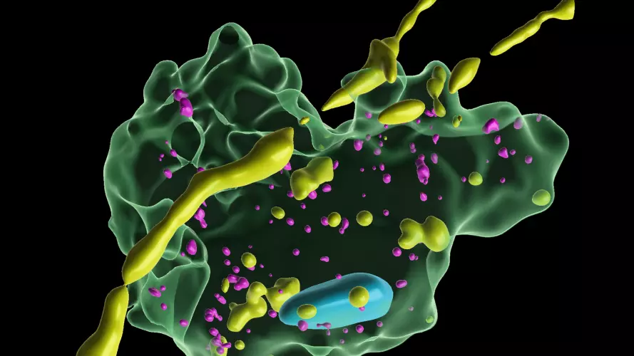
Kavli Oxford researchers discover how brain immune cells protect neurons by ‘nibbling’ away harmful material
13 October 2026
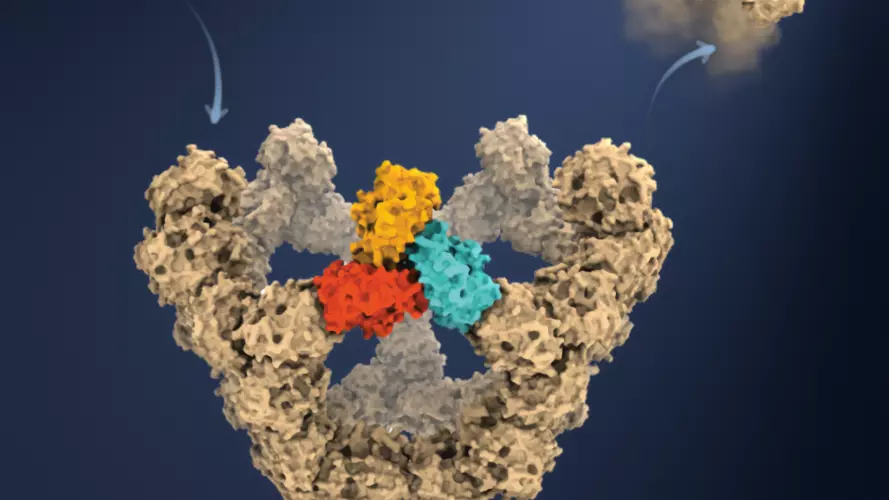
Kavli Oxford researchers capture the step-by-step assembly of individual virus-like particles
16 September 2026
Kavli Oxford researchers receive Wellcome Early-Career Awards
14 September 2026
Pagination
Previous page
Previous
Current page
1
Page
2
Page
3
Page
4
Page
5
Page
6
Page
7
…
Page
48
Next page
Next